Petroclival Meningioma: Posterior Petrosal Approach
This is a preview. Check to see if you have access to the full video. Check access
Complex Petroclival Meningioma: Posterior and Anterior Petrosectomies
Petroclival meningiomas are rare and account for less than 2% of all meningiomas. These tumors arise along the upper two-thirds of the clivus, superior to the jugular foramen and medial to the cranial nerves’ foramina at the petroclival junction. This anatomic location is uniquely difficult to approach surgically because it is very closely surrounded by multiple neurovascular structures, including the brainstem, basilar artery, cranial nerves (CNs) III-VIII, the cavernous sinus, and the sella.
This location also resides at the convergence of the supra- and infratentorial compartments, allowing petroclival meningiomas to span both spaces. This anatomic configuration may require radical skull base osteotomies or combined/staged approaches. Tumors of the lower one-third of the clivus, inferior to the jugular foramen, are primarily foramen magnum lesions and are discussed in the chapter dedicated to the Foramen Magnum Meningioma.
The natural history of petroclival meningiomas is presumed to be similar to that of other meningiomas, but it has not been well documented because of the relative rarity of these lesions. In some cases these tumors show evidence of slow, steady growth, but in others, growth is sporadic. Some lesions are followed unchanged for many years until they suddenly begin to enlarge, whereas others enlarge initially then stop growing. Some of these lesions never manifest any evidence of growth. Petroclival meningiomas often grow quite large before diagnosis, further contributing to the difficulty of their resection.
As they grow, petroclival meningiomas displace the brainstem posteriorly and toward the contralateral side, becoming intimately involved with the distal basilar artery, its branches, and perforators. The lateral aspect of the tumor is often draped by thinly splayed cranial nerves lying between the surgeon and the tumor. Cavernous sinus invasion is common.
The complex anatomy of the adjacent temporal bone creates a surgical lesion that is surrounded by vulnerable structures and difficult to expose. Although the development of microsurgical techniques, skull base approaches, improved imaging, and stereotactic radiosurgery have all contributed to decreasing morbidity rates over the past few decades, resection of petroclival meningiomas remains a formidable surgical challenge to even the most experienced skull base surgeons.
Clinical Presentation
As with other meningiomas, petroclival meningiomas most commonly affect middle-aged women. The clinical course is insidious, with subtle onset of cranial nerve, cerebellar, and brainstem dysfunction.
Presenting symptoms are usually a result of cranial nerve compression with CN V most frequently involved. Patients often present with headaches, facial pain or numbness, diplopia, hearing loss, vertigo, facial weakness, dysphonia, or dysphagia.
Loss of coordination and gait problems are also frequent complaints. Physical examination often reveals one or more cranial neuropathies and long tract signs and cerebellar ataxia.
Evaluation
Following a thorough patient history and physical examination with particular attention to the cranial nerves, cerebellar, and long tract function, MR imaging with and without gadolinium enhancement is in order. Preoperative auditory testing is performed to assess for useful hearing. This will influence the choice of surgical corridor.
The characteristics of the tumor are evaluated, including size, location, and the presence of calcification. The tumor’s relationship to the brainstem, cranial nerves, temporal bone, cavernous sinus, and vasculature are also assessed. Edema within the brainstem is an ominous sign and signifies the loss of arachnoid planes and presence of pial invasion. This feature drastically increases the risk of surgical intervention.
A CT scan identifies tumor calcification and hyperostosis of the adjacent temporal bone. Imaging of the vasculature is helpful to better assess displacement or encasement of vascular structures by the tumor, the patency of venous sinuses, and location of temporal venous drainage such as the vein of Labbé. The route of this vein can influence the choice of surgical approach.
In the case of cavernous sinus involvement or unilateral vertebral artery encasement, a balloon occlusion test determines the feasibility of arterial sacrifice with or without the need for bypass. I do not believe vascular sacrifice and revascularization are indicated for benign meningiomas in an era when radiosurgery can effectively control small residual tumors.
Embolization of these tumors is usually not beneficial because cannulating the meningohypophyseal trunk is technically difficult and fraught with risks. If a petrosal approach is used, devascularization occurs via extradural removal of bone during surgical exposure.
Other considerations for protecting vital venous structures cannot be overstated. Dynamic CT angiograms and venograms are especially worthwhile. Large caliber veins and other relevant vascular structures encountered during the approach, such as crossing veins during a craniotomy and tentorial transectioning, should be protected. More specifically, the posterior location of the vein of Labbé (traversing through the tentorium) and the presence of large draining veins within the tentorium can persuade me to avoid the anterior or posterior petrosal approach and consider staged or combined posterior fossa and pterional craniotomies.
Although the basilar artery appears to be encased in many cases, intact arachnoid planes that allow safe dissection of the artery can usually be developed during surgery.
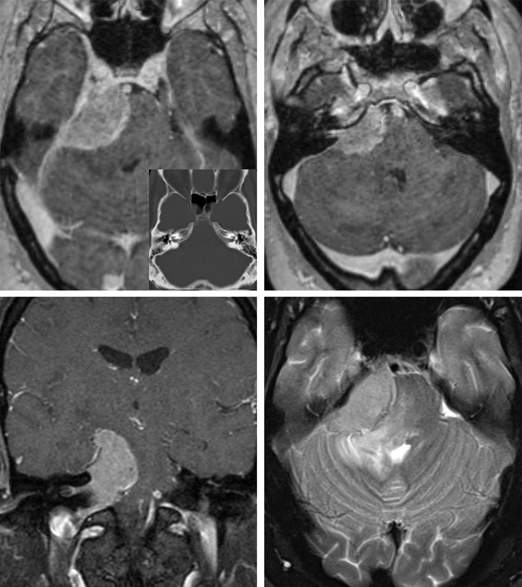
Figure 1: A large petroclival meningioma favorable for the posterior petrosal approach is shown. The tumor extends inferior to CN VII/VIII complex. Note infiltration of the VII/VIII foramen by the tumor. Presence of edema in the brainstem and middle cerebellar peduncle is worrisome for absence of intact pial planes during microsurgery.
Indications for Surgery
Once the patient and all studies have been thoroughly evaluated and the results discussed with the patient and family, a treatment plan can be devised. Treatment options for petroclival meningiomas include surgical resection, stereotactic radiosurgery, and observation with serial imaging.
Indications for treatment include neurologic deficit from tumor mass effect and documented tumor growth shown on serial imaging studies. The best chance for cure of a meningioma is gross total resection with removal of the involved dura and bone. The initial surgery is the patient’s best chance for cure; however, aggressive efforts for gross total resection of a petroclival meningioma often result in permanent neurologic morbidity. Brainstem perforating vessels, draining veins, and vascular supply to cranial nerves are particularly vulnerable, and damage to these structures often goes unnoticed during surgery.
The surgical goal is maximal safe resection. The practice of leaving residual tumor on vulnerable neurovascular structures in order to maintain neurologic function is highly advised. Residual tumor can be observed via serial MRIs or treated with stereotactic radiosurgery (SRS). This combination of adaptive surgery and SRS has emerged as a new treatment paradigm. Some neurosurgeons plan subtotal resection for tumors that are known to involve delicate neurovascular structures (i.e., arterial encasement seen on imaging). Heavy calcification also correlates with a high risk of tumor encasement and adherence.
For smaller asymptomatic tumors or minimally symptomatic tumors in high surgical risk patients, SRS may be used as the primary treatment modality.
Selection of the Surgical Approach for Petroclival Meningioma
When determining the best approach for a specific petroclival meningioma, I classify the tumor based on its location along the clivus and in relation to the cranial nerve foramina:
- Lesions confined to the upper one-third of the clivus.
- Extension lateral to the internal auditory canal (IAC)
- Suprasellar or parasellar extension
- Lesions confined to the middle one-third of the clivus
- Extension across the midline anteriorly
- Lesions confined to the posterior fossa with primarily lateral extension
- Lesions involving two or more of the above classifications.
Petroclival meningiomas may be approached through several different operative corridors, and selection of the “ideal” surgical approach requires a detailed analysis of the lesion in relation to anatomic landmarks.
For lesions confined to the upper one-third of the clivus (group 1, above), an anterior petrosal approach is appropriate through a pterional or subtemporal craniotomy. The medial exposure of this approach is adequate for tumors that extend to the midline and slightly beyond, but it is limited laterally by the IAC.
Tumors with extension lateral and inferior to the IAC (group 1a, above) will require a posterior petrosectomy or retrosigmoid approach. Tumors with a significant suprasellar or parasellar component (group 1b, above) are reached through an orbitozygomatic (OZ) or pterional craniotomy. The OZ approach is combined with the anterior petrosectomy for select masses to address the petroclival component. Giant tumors requiring both an OZ and posterior petrosal approach are staged for two reasons: 1) head positioning cannot accommodate these two approaches in combination, and 2) long operative sessions should be avoided.
For lesions within the middle third of the clivus (group 2, above), I recommend the posterior petrosal route. This route combines a posterior petrosectomy with subtemporal and suboccipital craniotomies. Posterior petrosal approaches provide varying degrees of exposure of the medial clivus and anterolateral brainstem depending on the degree of petrous pyramid resection. This osteotomy varies from a partial mastoidectomy and exposure of the Trautmann triangle to a transcochlear petrosectomy during which the surgeon removes the entire otic capsule and mobilizes the facial nerve. The latter radical approach is not necessary.
The patient’s preoperative hearing and facial nerve function determine the extent of petrous apex osteotomy. Importantly, a high-riding jugular bulb restricts the inferior extent of this exposure. Furthermore, the anatomic variations near the vein of Labbé may prohibit division of the superior petrosal sinus, further limiting the utility of this approach.
If the patient’s hearing is intact, a less radical posterior petrosectomy (retrolabyrinthine) is used for lesions extending beyond the midline (group 2a, above). I do not believe a more extensive bone resection is needed because tumor debulking provides an ample amount of space to reach the anterior brainstem and the contralateral side.
The retrosigmoid approach is appropriate for petroclival lesions confined to the infratentorial compartment with primarily lateral extension from their petroclival origin (group 3, above). This workhorse approach is associated with less morbidity than other skull base approaches. Although this approach typically may not access the anterolateral brainstem, petroclival meningiomas displace the brainstem posteriorly and contralaterally, potentially creating an adequate surgical corridor.
The suprameatal tubercle can be drilled and the tentorium divided to widen the corridor anteriorly and superiorly, but significant tumor extension into the middle fossa or anterior to the dorsum sella cannot be addressed through the retrosigmoid approach. In many cases, this transtentorial modification is a long reach and may not provide adequate exposure without significant retraction on the cerebellum.
Tumors that occupy more than one of the above anatomic compartments are resected via combined/staged approaches. Lastly, as endoscopic endonasal techniques continue to improve, small petroclival meningiomas may prove to be resectable in this manner because early devascularization of the tumor is achieved through a corridor medial to the involved cranial nerves, allowing the surgeon to encounter the tumor before the neural tissue.
For more information, please refer to the Anterior Petrosectomy and Conservative Posterior Petrosectomy chapters.
I do believe that the extended retromastoid approach is adequate for removal of petroclival meningiomas that do not have a significant supratentorial component. This approach avoids the risk of morbidity associated with petrosal osteotomies with no proven compromise in exposure and tumor resection.
Operative Anatomy
The anatomy of the petroclival region is complex. Detailed study of the region in the microsurgical laboratory is mandatory.
Click here to view the interactive module and related content for this image.
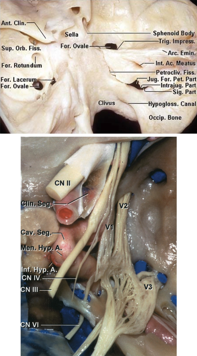
Figure 2: Posterior oblique view of the clivus is shown. Note the location of the petroclival fissure from which most petroclival meningiomas arise (top photo). The relevant neurovascular anatomy on the right side is also included (bottom photo). Cranial nerve VI travels under the dura along the clivus before entering the cavernous sinus. Coagulation in this part of the clival dura can lead to the nerve’s permanent injury. (Images courtesy of AL Rhoton, Jr.)
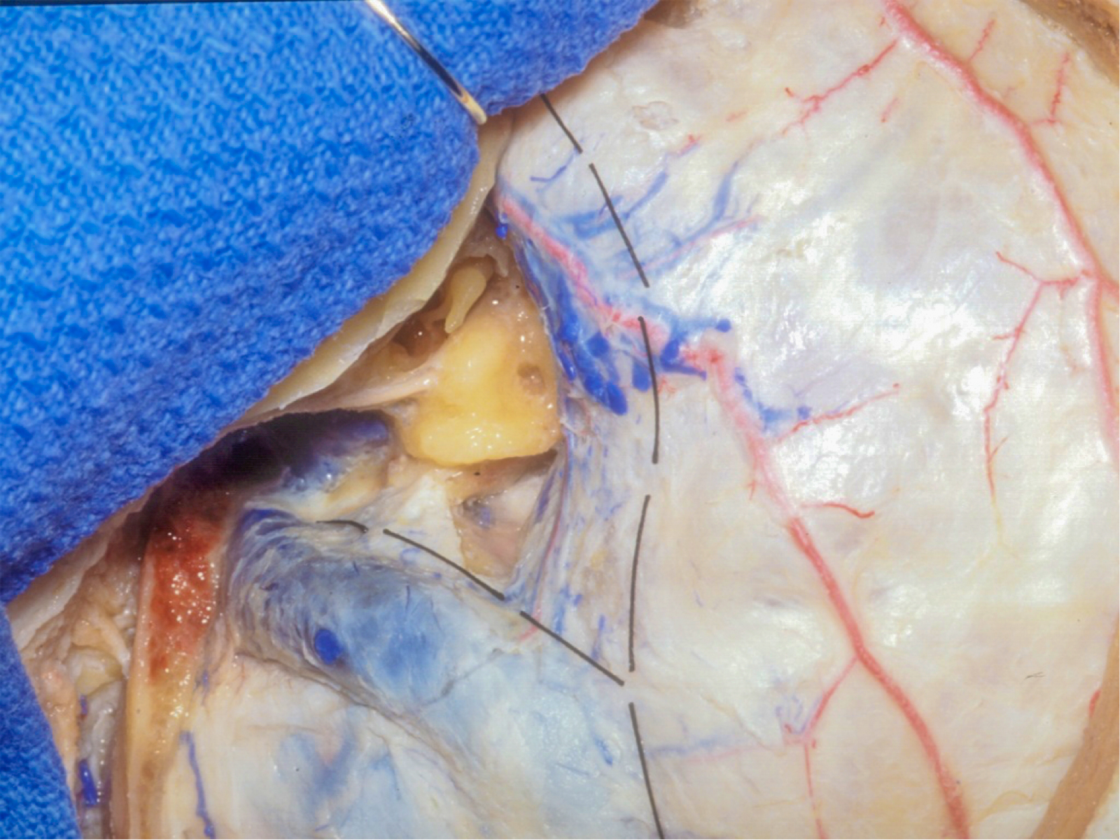
Figure 3: Osteotomy and dural incisions for a posterior petrosal osteotomy are demonstrated. (Courtesy of AL Rhoton, Jr.)
Figure 4: The neurovascular exposure following completion of the dural opening and tentorial incision. Note the panoramic view of the petroclival cisterns (top image). Mobilization of the temporal lobe can further expand the subtemporal operative trajectory (bottom image). Retrolabyrinthine presigmoid exposure allows preservation of the semicircular canals. (Courtesy of AL Rhoton, Jr.)
Figure 5: If necessary, the semicircular canals and vestibule can also be removed to complete the translabyrinthine approach. However, this approach is rarely necessary. (Courtesy of AL Rhoton, Jr.)
RESECTION OF PETROCLIVAL MENINGIOMA: POSTERIOR PETROSAL APPROACH
Before positioning the patient for surgery, I routinely place a lumbar drain to facilitate extradural drilling and exposure via dural sac decompression. This is an important step for petrosal bone drilling. Please see the Extended Posterior Petrosectomy chapter for more details regarding exposure.
Neurophysiologic monitoring is recommended. Facial nerve, brainstem auditory evoked responses (BAER), and somatosensory evoked potentials (SSEP), and (when necessary) lower cranial nerves are commonly monitored.
INTRADURAL PROCEDURE
Since the posterior petrosal approach is usually used for petroclival meningiomas, the intradural procedure for this exposure is primarily discussed herein. Note that very similar intradural dissection principles apply when using the anterior petrosal route. The resection techniques for removal of petroclival meningiomas through the anterior petrosal approach are discussed in the Petroclival Meningioma: Anterior Petrosectomy chapter.
If there is a large infratentorial component of the tumor, a second dural opening posteroinferior to the transverse-sigmoid junction can be created to expand the working angles into the cerebellopontine cisterns.
Upon opening the arachnoid bands around the tumor, the cerebellum falls away naturally. The temporal lobe may be gently mobilized superiorly and held in place using fixed retractors. Neurovascular structures may be invested or stretched in and/or around the mass, so dissection must be performed carefully to identify neurovascular structures early and keep them out of harm’s way during the later stages of the operation.
Devascularization begins with coagulating the tumor’s insertion at the dura overlying the petrous bone and meningeal feeders from the tentorium. Debulking proceeds in a piecemeal fashion via a combination of bipolar emulsification/suction evacuation and ultrasonic aspiration. The ultrasonic aspirator is quite effective for operating on fibrous tumors while avoiding traction on the neural structures during manipulation of the tumor capsule.
The surgeon takes care to preserve the cranial nerves (CNs) and arteries that may be embedded in the tumor bulk. Rootlets from CN V may be splayed over the anterior surface of the tumor, whereas CNs VII and VIII are usually displaced posteroinferiorly or embedded within the tumor. Some of the rootlets fo CN V may not be salvagable because of their adherence to the tumor capsule, however; most rootlets must be preserved to avoid excessvie neuropathy/numbness. Cranial nerve VI is displaced anteriorly and inferiorly; it is most at risk because of its small diameter and long intracisternal course.
Figure 6: Although the trigeminal nerve is often lateral to these tumors as the base of the tumor is medial to this nerve, it is not unusual for the large tumors to extent medial, over and lateral to the nerve, displacing the nerve inferiorly. The tentorial incision is continued anteriorly over the trigeminal nerve so that the Meckel's cave and the posterior cavernous sinus are unroofed. The part of the tumor in the Meckel's cave is usually easily dissectable from the nerve. It is critical that the base of the tumor medial to the trigeminal nerve is heavily coagulated in order to devascularize the tumor. Early release of the trigeminal nerve within the cave is advantageous to avoid undue traction on the nerve. This nerve is at the center of the exposure and its manipulation is unavoidable to handle the tumor bulk medial to the nerve.
Figure 7: After aggressive devascularization of the tumor at its base along the superior clival dura and tentorium, an ultrasonic aspirator is used to debulk the tumor in expectation of its dissection from the CN VII-VIII complex and trigeminal nerve. At least, one fixed retractor is needed to hold the temporal lobe in place.
Figure 8: The tumor capsule is then microsurgically and sharply dissected from the medial aspect of the CN VII-VIII complex.
Figure 9: Cycles of debulking and microsurgical capsular dissection continue until the bulk of the tumor is dissected and reduced from the upper brainstem. The trochlear nerve is often incorporated into the tumor capsule and its salvage is not possible. I have lost this nerve during dissection in certain patients; despite this loss, some of these patients did not suffer from a trochlear nerve palsy. It appears that the chronic compression of this very fine nerve allows for compensatory control of the superior oblique muscle by the other ocular muscles. The posterior cerebral and superior cerebellar arteries may be engulfed.
Figure 10: The last piece of the tumor is delivered out of the Meckel's cave. The perforating vessels in this area and to the diencephalon can be easily injured despite their delicate handling. Their spasm can lead to devastating consequences including thalamic infarction. I use papaverine soaked pledgets to periodically bathe the perforators during the entire dissection process.
Figure 11: This final illustration demonstrates some of the displacement patterns of cranial nerves. The oculomotor nerve is along the superior aspect of the capsule. The affected part of the dura is coagulated to minimize the risk of recurrence.
Aggressive tumor debulking followed by meticulous microsurgical mobilization of the thin/flexible tumor capsule away from the cranial nerves is critical. The surgeon should not overlook the edges of the tumor capsule that guide the planes of dissection. The perforating vessels to the cranial nerves should also be protected. The labyrinthine artery should be preserved to minimize the risk of hearing loss. Indiscriminate pulling and traction on the capsule along the operative blind spots leads to vascular avulsion injuries. The encased anterior inferior and posterior inferior cerebellar arteries are identified and protected.
After debulking the tumor, the operator’s attention alternates between the supratentorial and infratentorial compartments in order to identify the cranial nerves and dissect them free from the tumor capsule. The nerves are not directly handled, but their encasing arachnoid bands are gently mobilized away from the tumor capsule. Please see more details about handling neurovascular structures in the chapter titled Lessons Learned of this volume.
I usually ask my assistant to simultaneously irrigate the operative field and use the suction device to keep the operative field clear while I use a pair of microforceps (such as jeweler's forceps) in each hand to tease away the tumor capsule from the blood vessels or cranial nerves.
The arachnoid planes should be preserved to avoid loss of function. If the pia of the brainstem is disrupted, aggressive tumor resection has disastrous consequences and subtotal tumor removal is mandatory. The basilar artery may be embedded or displaced to the contralateral side; its branches are susceptible to injury during tumor dissection. Tumor infiltration of the cranial nerves’ foramina minimizes the chance of gross total resection because the risk of cranial nerve injury during manipulation of the tumor within the foramina is substantial.
Although the first operation is the patient’s best chance for a surgical cure, it is wise to leave adherent tumor attached to delicate structures if the arachnoid planes have been lost.
The infiltrated dura is coagulated to minimize the risk of future tumor recurrence. Thermal injury to the cranial nerves must be avoided by using copious irrigation.
Closure
Once the surgical goals have been achieved, the surgeon’s attention turns to closure. Primary dural closure is impossible and alternative methods are applied. The available dural flaps are approximated.
Adipose tissue with its globular texture is one of the best barriers against cerebrospinal fluid leakage. Strips of adipose tissue are placed across the dural opening to seal the dural defect. Before placement of the adipose grafts, all exposed air cells are meticulously waxed.
Alternatively, a vascularized tissue flap prepared during the exposure may be rotated to fill the defect in the dura. This method is used for cases involving repeat operations or when the patient has a prior history of radiation treatment. The periosteal flap is draped over the drilled petrous surface. The bone flap is replaced and secured using miniplates, and the rest of closure is conducted in a standard fashion. A mastoid head dressing can potentially decrease the risk of pseudomeningocele formation.
A lumbar drain is used to drain 8 cc per hour of cerebrospinal fluid for 48 hours after surgery. Patients are mobilized as soon as possible.
Pearls and Pitfalls
- Petroclival meningiomas are notoriously challenging tumors and their intrinsic features (density, viscosity, and tenacious adherence to surrounding structures) may render complete resection prohibitively dangerous independent of the surgeon’s skill level.
- The important factors facilitating resection of these tumors relates to 1) intact arachnoid planes separating the tumor capsule from neurovascular structures, 2) the texture/consistency of the tumor, and 3) adherence of the tumor to the basilar artery and its perforators.
For additional illustrations of the combined approach craniotomy for posterior and middle fossa access, please refer to the Jackler Atlas by clicking on the image below:
Contributors: Andrew R. Conger, MD, MS, and Stephen Grupke, MD, MS
References
Al-Mefty O. Meningiomas. New York: Raven Press, 1991.
Al-Mefty O. Operative Atlas of Meningiomas. Philadelphia: Lippincott-Raven, 1998.
Cho C, Al-Mefty O. Combined petrosal approach to petroclival meningiomas. Neurosurgery. 2002;51:708-718.
Coppens J, Couldwell W. Clival and petroclival meningiomas, in Demonte F, McDermott M, Al-Mefty O (eds): Al-Mefty’s Meningiomas, 2nd ed. New York: Thieme Medical Publishers, 2011, 270-282.
Liu J, Couldwell W. Petrosal approach for resection of petroclival meningiomas, in Badie B (ed): Neurosurgical Operative Atlas: Neuro-Oncology, 2nd ed. Rolling Meadows, IL: Thieme Medical Publishers and the American Association of Neurological Surgeons, 2007, 170-179.
Rhoton AL Jr. The cavernous sinus, the cavernous venous plexus, and the carotid collar. Neurosurgery. 2002; 51(4 Suppl) S1-375-410.
Starke R, Kano H, Ding D, et al. Stereotactic radiosurgery of petroclival meningiomas: a multicenter study. J Neurooncol. 2014;119:169-176.
Tew JM, van Loveren HR, Keller JT. Atlas of Operative Microneurosurgery. Philadelphia: Saunders, 1994-2001.
Xu F, Karampelas I, Megerian CA, Selman WR, Bambakidis NC. Petroclival meningiomas: an update on surgical approaches, decision-making, and treatment results. Neurosurg Focus. 2013;35:E11.
Please login to post a comment.


