Pituitary Macroadenoma
Figure 1: Harvey Cushing abandoned transsphenoidal surgery in approximately 1929 as his transcranial techniques progressed and were deemed safe. The transcranial route allowed him to more thoroughly decompress the chiasm. In these photos, Cushing demonstrated his classic “frontal” incision for resection of pituitary adenomas.
Please note the relevant information for patients suffering from pituitary adenoma is presented in another chapter. Please click here for patient-related content.
This is a preview. Check to see if you have access to the full video. Check access
Endoscopic Transsphenoidal Removal of a Giant Pituitary Adenoma
Diagnosis, Indications for Surgery and Preoperative Considerations
For a detailed discussion of the diagnostic indications for surgery and preoperative considerations in patients with suspected pituitary adenoma, please refer to the chapter on Pituitary Adenoma: Diagnosis and Operative Considerations.
Figure 2: Pituitary macroadenomas are larger than 10 mm in diameter and demonstrate mass effect on the surrounding structures. Their compression is most clinically evident as bitemporal hemianopsia from mass effect on the chiasm and/or optic nerves.
Figure 3: The configuration of a typical pituitary adenoma on a sagittal plane is shown here. Note the location of the compressed pituitary gland in the posterior or superior directions.
PTERIONAL CRANIOTOMY FOR RESECTION OF PITUITARY ADENOMAS
Use of the pterional approach for pituitary adenoma resection is exceedingly rarely indicated. In less than 3% of patients, I use the transcranial, and more specifically, the pterional approach for reaching residual tumors unattainable through transsphenoidal surgery.
The major indications for the transcranial approach include significant intracranial tumor expansion well beyond the parasellar boundaries and a dumbbell-shaped tumor with a very narrow diaphragmatic neck. The presence of ectatic carotid arteries projecting along the midline is another contraindication to the transsphenoidal route, justifying the use of transcranial corridor.
The major limitations of the transcranial approach are the need for brain manipulation and difficulty in accessing the intrasellar component of the tumor, which is conveniently accessed transsphenoidally. The use of an endoscope has expanded the reach of the transsphenoidal route, obviating the need to use a transcranial corridor in almost all cases.
The remainder of this section focuses on nuances of technique for using the pterional route for reaching and resecting pituitary adenomas. For additional technical guidance for performing the pterional approach, please refer to the chapter on Pterional Craniotomy.
After completion of the craniotomy and dural opening, the anterior Sylvian fissure is split and the frontal lobe gently mobilized using dynamic retraction. The regional arachnoid membranes are dissected open and the parachiasmatic area is widely exposed. The tumor is typically covered by the ipsilateral optic nerve and the diaphragm encases the superior tumor capsule. The ipsilateral optic nerve can be relieved by large-scale decompression via operative trajectories medial and lateral to the nerve.
The lateral trajectory requires dissection between the carotid artery and the optic nerve. The internal carotid artery perforating vessels on the inferior and posterior surface of the optic chiasm and their associated arachnoid layers should be carefully protected. Preservation of these perforators decreases the risk of hemorrhage and ischemic infarction of the surrounding vital tissues; these complications are a significant source of morbidity from this operation.
An adherent tumor capsule poses a significant surgical challenge. If the capsule is attached to the perforating vessels of the carotid artery, posterior communicating artery, or middle cerebral artery, subtotal resection is prudent. The conservative approach involves removing only the core of the macroadenoma and leaving the capsule behind to preserve the tethered critical structures.
Adherent tumor capsule often engulfs the small and medium-size arteries at the skull base and therefore is refractory to total excision. The extra risk imposed by an attempted radical capsule removal is not justified. In this setting, the surgeon should focus on decompression of the nerves/chiasm and permissible adenoma resection, not endangering other structures. Radiotherapy is a reasonable option after partial resection.
Figure 4: This young patient with a giant off-midline asymmetrical macroadenoma underwent surgery via an expanded transnasal endoscopic approach (upper images). The lateral and posterior parasellar components were not easily resectable through the transnasal route because of the tumor’s consistency and a “blind” lateral operative reach. Suprasellar tumor decompression was accomplished, but significant postoperative residual tumor continued to cause mild optic apparatus and brainstem compression, limiting the efficacy of radiosurgical therapy (lower images). A pterional craniotomy was deemed appropriate to further decompress the optic nerve and brainstem. During the second surgery, the tumor was debulked and the nerves decompressed but capsule was found to be very adherent and was left behind to protect the perforating arteries.
Transcranial Approach to Pituitary Adenoma
TRANSNASAL TRANSPHENOIDAL RESECTION OF PITUITARY MACROADENOMAS
This procedure begins with standard preparation for transsphenoidal surgery with a noncaustic antiseptic agent used to cleanse the patient’s nasal compartment, gingival surface, and lower face. Placement of intranasal absorbable cotton soaked with a dilute cocaine solution is optionally used to induce vasospasm within the mucosa. This promotes hemostasis during the initial submucosal dissection.
I previously discussed the technical nuances for the microscope-guided approach in the chapter on Microscope-Guided Endonasal Transsphenoidal Approach of the Cranial Approaches volume. Moreover, the endoscopic-guided route is discussed in the Endoscopic Expanded Transnasal Approach. Please refer to these chapters for information regarding the initial and final stages of the operation, including exposure and closure.
The techniques of macroadenoma resection during microscope- and endoscope-guided surgeries are very similar and will be discussed herein. The endoscopic method offers significantly enhanced direct visualization of the operative space and greater working angles, thereby minimizing the risk of an inadvertent injury to the surrounding structures.
Specifically, macroadenoma resection is more efficient under endoscopic guidance because of the importance of wide visualization that increases the operator’s confidence in more aggressive maneuvers and resection. In other words, the improved view of the lateral blind spots via angled endoscopes promotes maneuvers that would have been considered risky under microscopic guidance.
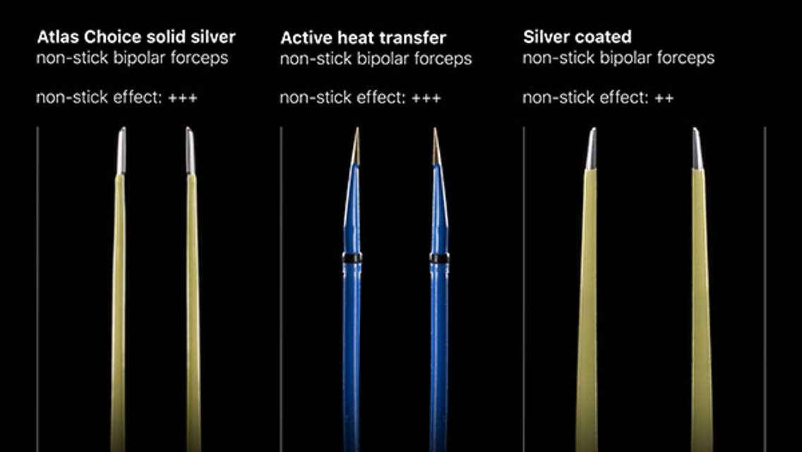
Figure 5: When wide bony removal at the sellar floor is complete, the exposed dura, including the intercavernous sinus, is gently cauterized using bipolar electrocautery. I use an ample amount of FLOSEAL hemostatic matrix (Baxter, Deerfield, IL) to seal the bleeding from the cavernous sinus and other venous lakes within the exposed dura. Overcoagulation of the dura leads to its shrinkage and exacerbation of epidural venous bleeding.
The principle of judicial minimalism in bone removal, while not compromising adequate lesional exposure, microsurgery, and tumor removal, is pertinent. The most common cause of subtotal resection is inadequate bone removal and resultant restricted dural opening. I create a wide bony opening along the floor of the sella, encompassing the walls of both carotid arteries.
Before dural opening, a circumferential bony edge should be mucosa-free to allow reconstruction of the floor at the end of the operation. Ultrasonography may be used to guide the dural opening and avoid injury to the carotid arteries. The dura is incised in a cruciate fashion, and a round dissector is used to widely dissect the tumor capsule from the inner surface of the dura. This maneuver expedites identification of the tumor capsule during the later stages of the operation when tumor debulking will make the capsule less identifiable (Redrawn from Tew, van Loveren, Keller*).
INTRADURAL PROCEDURE
The following section describes resection of macroadenoma through both microscope and endoscope-assisted methods. For a more detailed discussion of the advantages and disadvantages of microscope-assisted versus endoscope-guided adenoma resection, see Pituitary Adenoma: Diagnosis and Operative Considerations. I routinely employ the endoscopic route for resection of pituitary adenomas and I believe it provides remarkable advantages for maximizing safe tumor removal.
Figure 6: The first step of macroadenoma resection involves a sequential debulking procedure. The inferior pole of the tumor is punctured and morcellized by a ring curette; suction removes the dislodged tissue. A standardized approach to tumor debulking is encouraged and should begin inferiorly, then posteriorly, laterally, and finally superiorly. This sequence prevents premature descent of the diaphragm and its interference with tumor manipulation. Early exposure and descent of the diaphragm can obstruct the operator’s view for radical tumor removal and places the diaphragm at risk of tearing.
The central portion of the tumor should be avoided early due to the risk of applying downward traction on the diaphragm, thereby masking the lateral aspects of the adenoma. The superior aspect of the tumor should be cautiously manipulated because of the risk of injuring the pituitary stalk (Redrawn from Tew, van Loveren, Keller*).
Figure 7: Debulking should continue into the lateral aspect of the tumor, ending at the superior segment. After generous debulking, the tumor capsule is gently peeled off from the walls of the cavernous sinus. The angled endoscope allows a more expanded view of my operative maneuvers so that I can dissect the tumor capsule away from the medial walls of the cavernous sinus under direct vision rather than blindly searching for the lateral poles of the tumor using ring curettes. The location of the normal pituitary is ghosted in orange in this illustration. Note the use of a bimanual dissection technique (Redrawn from Tew, van Loveren, Keller*).
Figure 8: The medial wall of the cavernous sinus should be scrutinized carefully to remove any remaining tumor. This is a crucial step; residual tumor will prevent the later descent of the diaphragm and radical tumor removal. The direct line of sight of the microscope will not allow adequate visualization of these lateral corners. Blind aggressive maneuvers can lead to an injury to the carotid artery. For a discussion regarding management of carotid artery injuries, refer to the chapter on Pituitary Adenoma: Diagnosis and Operative Considerations.
In the event that the tumor invades the medial cavernous sinus wall or the diaphragm, intracavernous and supradiaphragmatic resection can be continued, but pursued cautiously. The endoscopic-expanded perception using angled endoscopes promotes conservative removal of tumor within the cavernous sinus under direct visualization (Redrawn from Tew, van Loveren, Keller*).
Figure 9: Upon resection of the infrasellar portion (especially the lateral components) of the adenoma, caudal prolapse of any suprasellar portion of the soft adenoma into the sellar compartment facilitates additional tumor removal. Fibrous or septated tumors (especially recurrent tumors) can resist this prolapse, making their removal challenging. Direct endoscopic visualization allows safe intrasellar sharp dissection of the septations.
In these situations, injection of 15 to 20 ml of lactated Ringer’s solution or air into the lumbar drain (placed preoperatively for giant tumors,) compressing the jugular veins bilaterally, and administering positive end-expiratory pressure can also assist with tumor mobilization. Patience is a virtue here as tumor descent is gradual. “Removing what the tumor gives you” rather than pulling indiscriminately on the blind corners is the safest strategy.
Following resection of this portion of the tumor, the diaphragm is visualized prolapsing into the sella. This is a good indication for adequate resection, but further inspection is necessary. If the diaphragm is not readily collapsing, there is usually residual tumor along the lateral aspects of the resection cavity preventing the descent of the diaphragm. Bony exposure may need to be expanded laterally and additional tumor removal secured in these regions. Blind reach is not recommended (Redrawn from Tew, van Loveren, Keller*).
Figure 10: The anterosuperior portions of the adenoma provide a challenge during their resection because of the risk of damaging the adjacent structures, most commonly the diaphragm. This is another common location for unintentional subtotal tumor removal; the residual tumor can continue to cause symptomatic optic apparatus compression. Angled endoscopes provide superior visualization in this region. The gland may be gently mobilized to expose residual tumor. Adequate bone resection is required to avoid residual tumor (Modified and Redrawn from Tew, van Loveren, Keller*).
Figure 11: A tear within the diaphragm and resultant cerebrospinal fluid leakage often occurs during blind forceful removal of a tumor located along the anterior aspect of the resection cavity above the tuberculum sella (anterior sellar recess) where the diaphragm is attached (inset image). It is important to identify the diaphragm and maintain its integrity throughout resection. If an inadvertent leak is apparent, I pack the previously prepared fat globules from the patient’s abdomen at the exact location of the leak and avoid indiscriminately and forcefully packing the entire sella because this maneuver may lead to suprasellar mass effect and vision impairment (Modified and Redrawn from Tew, van Loveren, Keller*).
Persistent bleeding from the resection cavity indicates residual tumor. The most appropriate technique for achieving hemostasis is gross total tumor removal. Upon removal of the last pieces of the tumor, bleeding immediately ceases spontaneously. Minimal packing of Gelfoam may be needed to stop venous bleeding.
After gross total resection of the tumor, the diaphragm will prolapse and essentially fill the entire sella. This prolapse can hide tumor fragments along the corners of the sella. To inspect these corners further, I place a small cottonoid patty on the diaphragm and gently mobilize the diaphragm superiorly using the suction device. This maneuver allows easy inspection of the gutters that frequently house additional pieces of tumor. Similarly, the collapsed folds of the diaphragm, often evident around giant tumors, conceal tumor and these hidden spots should be inspected carefully to avoid surprise findings on postoperative imaging.
The intact pituitary gland is more firm and vascular and should not be aggressively manipulated.
Formation of scar within the recurrent tumors prevents them from descending into the sella. More aggressive and nontraditional maneuvers including sharp dissection within the sella under careful endoscopic vision are often necessary to mobilize the tumor within septations and achieve reasonable resection. The risk of CSF leakage is high but this risk should not prevent radical subtotal removal. Intraoperative MRI has a role for advancing surgery of the recurrent tumors.
Fibrous Hypervascular Pituitary Macroadenoma: Facing a Daunting Challenge
Closure
Figure 12: I use abdominal fat graft for packing the sella after transsphenoidal surgery. The fat globules are wrapped in pieces of Surgicel to create spheres. This method facilitates their handling and directs their insertion/placement at the exact site of the leak. If Valsalva maneuvers exclude a leak, I still gently pack the sella to seal an occult leak. A piece of bony septum or prosthesis is used to keep the fat in place and reconstruct the sella floor. See the epidural location of the bone or prosthesis in the above illustration.
If a large defect in the diaphragm is created and the suprasellar contents are visible, a nasoseptal pedicled mucosal flap is prepared, and reconstruction is performed according to the procedure described in the chapter on Endoscopic Expanded Transnasal Approach (Modified Redrawn from Tew, van Loveren, Keller*).
Growth hormone and adrenocorticotropin-secreting macroadenomas present special challenges in their resection because their invasion of the cavernous sinus and surrounding dural membranes precludes their gross total resection in some cases. Patients harboring such tumors should undergo meticulous microsurgical resection to establish a cure. I pursue intracavernous removal of these tumors under endoscopic guidance. Transdiaphragmatic protrusion of these tumors requires sharp dissection of the diaphragm and their gross total removal.
Cushing's Disease and Macroadenoma: The Importance of the Transdiaphragmatic Reach
Postoperative Considerations
For a more detailed description of postoperative considerations, remission parameters for secreting tumors, and complications related to macroadenoma resection, see the chapter on Pituitary Adenoma: Diagnosis and Operative Considerations.
Pearls and Pitfalls
- Tumor resection along the medial aspects of the cavernous sinus may lead to venous bleeding. Cauterization in this region is not advised, but use of thrombin-soaked Gelfoam powder to pack the bleeding site is reasonable.
- Inadequate bone removal along the floor of the sella (frequently laterally or anteriorly) is the most common cause of inadequate tumor resection.
- Subtotal tumor removal and apoplexy of the residual tumor is typically the cause of symptomatic postoperative hematoma.
Contributor: Benjamin K. Hendricks, MD
For additional illustrations of using endoscopes during skull base surgery, please refer to the Jackler Atlas by clicking on the image below:
*Redrawn with permission from Tew JM, van Loveren HR, Keller JT. Atlas of Operative Microneurosurgery, WB Saunders, 2001. © Mayfield Clinic
Please login to post a comment.


