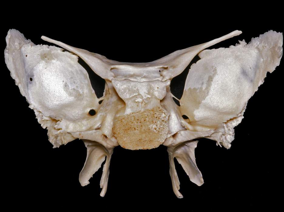
Posterior View of the Sphenoid Bone
5555
Surgical Correlation
Tags
Posterior view of an isolated sphenoid bone. It is the principal bone of the middle cranial fossa. It consists of a midline body, a pair of lesser wings that contain the optic canals, a pair of greater wings that contain a "crescent of foramina" including the superior orbital fissure, foramen rotundum, foramen ovale, and foramen spinosum, and a pair of pterygoid processes that project inferiorly. The pterygoid canals are located within the base of the pterygoid processes inferomedial to the foramen rotundum. The lesser wing articulates anteriorly with the frontal bone and cribriform plate of the ethmoid. The occipitosphenoid suture/synchondrosis marks the articulation between the dorsum sella and basilar portion of the occipital bone. Together, these form the clivus or the slope of bone anterior to the foramen magnum. (Image courtesy of AL Rhoton, Jr.)